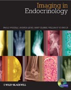
Imaging in Endocrinology
Pozzilli, Paolo
Lenzi, Andrea
Clarke, Bart L.
Jr., Young, William F.
Imaging in Endocrinology will provide endocrinologists and radiologists of all levels with an outstanding diagnostic imaging atlas to aid them in the diagnosis and management of all the major endocrine diseases they are likely to encounter. In full colour throughout, the 300 high–quality images consist of CT scans, MRI, NMR and histopathology slides, and are arranged by each specific endocrine condition, resulting in a visually outstanding and easily accessible tool that guides the user through exactly what to look out for and provides a practical and extremely useful aid in helping them formulate a diagnosis. Every major endocrine condition is covered in a specific section, including diseases of the thyroid, pituitary, reproductive and adrenal glands, the pancreas, bone metabolism problems, and the various forms of endocrine cancers. Each disease covered will offer a comparison of the normal findings so as to further assist in diagnosis. An accompanying website contains an online slide–atlas of all the figures in the book, to allow users to download all figures for use in presentations. Led by Paolo Pozzilli, an internationally–recognised expert in this field, the authors have assembled a wonderful collection of images that will be greatly valued by endocrinologists and radiologists alike, ensuring this is the perfect tool to consult when assessing patients with endocrine disease. INDICE: About the Companion Website, xii Preface, xiii Collaborators, xiv Chapter 1 Thyroid, 1 Hashimoto’s thyroiditis (chronic autoimmune thyroiditis), 1 Definition and epidemiology, 1 Etiology and pathogenesis, 1 Signs and symptoms, 1 Diagnosis, 1 Treatment, 1 Illustrations (Figs 1.1–1.4), 2–3 Graves’ disease (Basedow’s disease), 4 Definition and epidemiology, 4 Etiology and pathogenesis, 4 Signs and symptoms, 4 Diagnosis, 4 Treatment, 4 Illustrations (Figs 1.5 & 1.6), 5 Subacute thyroiditis (de Quervain’s thyroiditis), 6 Definition and epidemiology, 6 Etiology and pathogenesis, 6 Signs and symptoms, 6 Diagnosis, 6 Treatment, 6 Illustration (Fig. 1.7), 7 Benign thyroid nodules, 8 Definition and epidemiology, 8 Etiology and pathogenesis, 8 Signs and symptoms, 8 Diagnosis, 8 Treatment, 8 Illustrations (Figs 1.8–1.11), 9–10 Thyroid cancer, 11 Definition and epidemiology, 11 Etiology and pathogenesis, 11 Signs and symptoms, 11 Diagnosis, 11 Treatment, 12 Illustrations (Figs 1.12–1.25), 13–21 Chapter 2 Pituitary Gland, 22 Craniopharyngioma, 22 Definition, 22 Etiology, 22 Signs and symptoms, 22 Diagnosis, 22 Treatment, 22 Illustrations (Figs 2.1–2.3), 23 Hypothalamic dysgerminoma, 24 Definition, 24 Etiology, 24 Signs and symptoms, 24 Diagnosis, 24 Treatment, 24 Illustrations (Figs 2.4 & 2.5), 24 Growth hormone–secreting pituitary tumor, 25 Definition, 25 Etiology, 25 Signs and symptoms, 25 Diagnosis, 25 Treatment, 25 Illustrations (Figs 2.6–2.10), 25–26 Prolactin–secreting pituitary tumor, 27 Definition, 27 Etiology, 27 Signs and symptoms, 27 Diagnosis, 27 Treatment, 27 Illustrations (Figs 2.11–2.13), 27–28 Corticotropin–secreting pituitary tumor (Cushing syndrome), 29 Definition, 29 Etiology, 29 Signs and symptoms, 29 Diagnosis, 29 Treatment, 29 Illustrations (Figs 2.14–2.16), 30 Nelson syndrome, 31 Definition, 31 Etiology, 31 Signs and symptoms, 31 Diagnosis, 31 Treatment, 31 Illustrations (Figs 2.17–2.20), 31–32 Clinically nonfunctioning pituitary tumors, 33 Definition, 33 Etiology, 33 Signs and symptoms, 33 Diagnosis, 33 Treatment, 33 Illustrations (Figs 2.21–2.23), 34 Primary pituitary carcinoma, 35 Definition, 35 Etiology, 35 Signs and symptoms, 35 Diagnosis, 35 Treatment, 35 Illustrations (Figs 2.24–2.27), 36 Pituitary cyst, 37 Definition, 37 Etiology, 37 Signs and symptoms, 37 Diagnosis, 37 Treatment, 37 Illustrations (Figs 2.28–2.31), 38–39 Nontumorous lesions of the pituitary gland and pituitary stalk, 40 Definition, 40 Etiology, 40 Signs and symptoms, 40 Diagnosis, 40 Treatment, 40 Illustrations (Figs 2.32–2.34), 41–42 Pituitary apoplexy, 43 Definition, 43 Etiology, 43 Signs and symptoms, 43 Diagnosis, 43 Treatment, 43 Illustrations (Figs 2.35–2.37), 43–44 Tumors metastatic to the pituitary, 45 Definition, 45 Etiology, 45 Signs and symptoms, 45 Diagnosis, 45 Treatment, 45 Illustrations (Figs 2.38 & 2.39), 46 Chapter 3 Adrenal Gland, 47 Adrenal incidentaloma, 47 Definition, 47 Etiology, 47 Signs and symptoms, 47 Diagnosis, 47 Treatment, 47 Illustrations (Figs 3.1–3.4), 48–49 Adrenocortical carcinoma, 50 Definition, 50 Etiology, 50 Signs and symptoms, 50 Diagnosis, 50 Treatment, 50 Illustrations (Figs 3.5–3.8), 51 Adrenal–dependent Cushing syndrome, 52 Definition, 52 Etiology, 52 Signs and symptoms, 52 Diagnosis, 52 Treatment, 52 Illustrations (Figs 3.9–3.14), 53–54 Classic congenital adrenal hyperplasia, 55 Definition, 55 Etiology, 55 Signs and symptoms, 55 Diagnosis, 55 Treatment, 56 Illustrations (Figs 3.15 & 3.16), 56–57 Primary aldosteronism, 58 Definition, 58 Etiology, 58 Signs and symptoms, 58 Diagnosis, 58 Treatment, 58 Illustrations (Figs 3.17 & 3.18), 59–60 Adrenal venous sampling, 61 Definition, 61 Background, 61 Procedure and keys to successful AVS, 61 Interpretation of AVS, 61 Illustrations (Figs 3.19 & 3.20), 62 Primary adrenal failure (Addison’s disease), 63 Definition, 63 Etiology, 63 Signs and symptoms, 63 Diagnosis, 63 Treatment, 63 Illustrations (Figs 3.21–3.23), 64 Pheochromocytoma, 65 Definition, 65 Etiology, 65 Signs and symptoms, 65 Diagnosis, 65 Treatment, 65 Illustrations (Figs 3.24–3.30), 66–67 Paraganglioma, 68 Definition, 68 Etiology, 68 Signs and symptoms, 68 Diagnosis, 68 Treatment, 68 Illustrations (Figs 3.31–3.35), 69–71 Adrenal infarction, 72 Definition, 72 Etiology, 72 Signs and symptoms, 72 Diagnosis, 72 Treatment, 72 Illustrations (Figs 3.36 & 3.37), 73 Tumors metastatic to the adrenal glands, 74 Definition, 74 Etiology, 74 Signs and symptoms, 74 Diagnosis, 74 Treatment, 74 Illustrations (Figs 3.38 & 3.39), 75 Chapter 4 Pancreas, 76 Diabetes, 76 Diabetes and cardiovascular disease, 76 Definition and pathogenesis, 76 Etiology, 76 Signs and symptoms, 76 Diagnosis, 77 Treatment, 77 Illustrations (Figs 4.1–4.5), 77–79 Skin diseases associated with diabetes, 80 Definition, 80 Etiology, 80 Signs and symptoms, 80 Diagnosis, 80 Treatment, 80 Illustrations (Figs 4.6 & 4.7), 80–81 Diabetic foot, 81 Definition, 81 Pathophysiology and etiology, 81 Signs and symptoms, 81 Diagnosis, 82 Treatment, 82 Illustrations (Figs 4.8–4.22), 82–86 Diabetic neuropathy, 87 Definition, 87 Etiology, 87 Diagnosis, 87 Treatment, 87 Illustrations (Figs 4.23 & 4.24), 88 Diabetic nephropathy, 89 Definition, 89 Etiology, 89 Signs and symptoms, 89 Diagnosis, 89 Prevention and treatment, 90 Illustrations (Figs 4.25–4.28), 90–91 Diabetic retinopathy, 92 Definition, 92 Etiology, 92 Signs and symptoms, 92 Diagnosis, 92 Treatment, 92 Illustrations (Figs 4.29–4.38), 93–94 Insulinoma, 95 Definition, 95 Etiology, 95 Signs and symptoms, 95 Diagnosis, 95 Treatment, 95 Illustrations (Figs 4.39–4.42), 95–96 Acute pancreatitis, 96 Definition, 96 Etiology, 96 Signs and symptoms, 96 Diagnosis, 96 Treatment, 97 Illustrations (Figs 4.43–4.45), 97 Chronic pancreatitis, 98 Definition, 98 Etiology, 98 Signs and symptoms, 98 Diagnosis, 98 Treatment, 98 Illustrations (Figs 4.46–4.48), 98–99 Chapter 5 Bone and Mineral Metabolism, 100 Osteoporosis, 100 Definition, 100 Etiology, 100 Signs and symptoms, 101 Diagnosis, 101 Treatment, 101 Illustrations (Figs 5.1–5.5), 101–102 Primary hyperparathyroidism, 103 Definition, 103 Etiology, 103 Signs and symptoms, 103 Diagnosis, 103 Treatment, 104 Illustrations (Figs 5.6–5.16), 105–108 Hypoparathyroidism, 108 Definition, 108 Etiology, 108 Signs and symptoms, 109 Diagnosis, 109 Treatment, 110 Illustrations (Figs 5.17–5.19), 110 Pseudohypoparathyroidism, 111 Definition, 111 Etiology, 111 Signs and symptoms, 111 Diagnosis, 111 Treatment, 112 Illustrations (Figs 5.20 & 5.21), 112 Paget’s disease of bone, 113 Definition, 113 Etiology, 113 Signs and symptoms, 113 Diagnosis, 114 Treatment, 114 Illustrations (Figs 5.22–5.28), 114–116 Osteomalacia, 116 Definition, 116 Etiology, 116 Signs and symptoms, 117 Diagnosis, 118 Treatment, 118 Illustrations (Figs 5.29–5.35), 119–120 Chronic kidney disease, 121 Definition, 121 Etiology, 121 Signs and symptoms, 121 Diagnosis, 121 Treatment, 122 Illustrations (Figs 5.36–5.40), 123–124 Fibrous dysplasia, 125 Definition, 125 Etiology, 125 Signs and symptoms, 125 Diagnosis, 126 Treatment, 126 Illustrations (Figs 5.41–5.47), 127–128 Selected sclerosing bone disorders, 129 Definition, 129 Osteopetrosis, 129 Etiology, 129 Signs and symptoms, 129 Diagnosis, 130 Treatment, 130 Illustrations (Figs 5.48–5.51), 131 Pycnodysostosis, 132 Etiology, 132 Signs and symptoms, 132 Diagnosis, 132 Treatment, 132 Illustrations (Figs 5.52–5.54), 133 Melorheostosis, 134 Etiology, 134 Signs and symptoms, 134 Diagnosis, 134 Treatment, 134 Illustrations (Figs 5.55 & 5.56), 134 Progressive diaphyseal dysplasia (Camurati–Engelmann disease), 135 Etiology, 135 Signs and symptoms, 135 Diagnosis, 135 Treatment, 135 Illustration (Fig. 5.57), 136 Hepatitis C–associated osteosclerosis, 136 Etiology, 136 Signs and symptoms, 136 Diagnosis, 136 Treatment, 136 Illustrations (Figs 5.58–5.60), 137 Familial high bone mass phenotype due to Lrp5 mutation, 138 Etiology, 138 Signs and symptoms, 138 Diagnosis, 138 Treatment, 138 Illustrations (Figs 5.61 & 5.62), 138 Erdheim–Chester disease, 139 Etiology, 139 Signs and symptoms, 139 Diagnosis, 139 Treatment, 139 Illustrations (Figs 5.63–5.65), 140 Skeletal fluorosis, 141 Etiology, 141 Signs and symptoms, 141 Diagnosis, 141 Treatment, 141 Illustrations (Figs 5.66 & 5.67), 142 Tumoral calcinosis, 143 Definition, 143 Etiology, 143 Signs and symptoms, 143 Diagnosis, 144 Treatment, 144 Illustrations (Figs 5.68 & 5.69), 144–145 Osteogenesis imperfecta, 145 Definition, 145 Etiology, 145 Signs and symptoms, 145 Diagnosis, 147 Treatment, 147 Illustrations (Figs 5.70–5.77), 148–149 Other skeletal disorders, 150 Osteoporosis–pseudoglioma (OPPG) syndrome, 150 Definition, 150 Etiology, 150 Signs and symptoms, 150 Diagnosis, 150 Treatment, 150 Illustrations (Figs 5.78–5.81), 150–151 Hajdu–Cheney syndrome (hereditary osteodysplasia with acro–osteolysis), 152 Definition, 152 Etiology, 152 Signs and symptoms, 152 Diagnosis, 152 Treatment, 152 Illustration (Fig. 5.82), 152 Hypophosphatasia, 153 Definition, 153 Etiology, 153 Signs and symptoms, 153 Diagnosis, 153 Treatment, 154 Illustrations (Figs 5.83 & 5.84), 154 Chapter 6 Gonads, 155 Male gonads, 155 Testes, 155 Definition, 155 Function, 155 Tests, 155 Treatment, 156 Illustrations (Figs 6.1–6.4), 156–157 Testicular cancer, 158 Definition and epidemiology, 158 Etiology, 158 Signs and symptoms, 158 Diagnosis, 158 Treatment, 159 Illustrations (Figs 6.5–6.8), 159–162 Cryptorchidism, 163 Definition, 163 Etiology, 163 Signs and symptoms, 163 Diagnosis, 163 Treatment, 163 Illustrations (Figs 6.9 & 6.10), 164–165 Gynecomastia, 166 Definition, 166 Etiology, 166 Diagnosis, 166 Treatment, 167 Illustrations (Figs 6.11–6.13), 167–168 Klinefelter syndrome, 169 Definition, 169 Etiology, 169 Signs and symptoms, 169 Diagnosis, 169 Treatment, 169 Illustrations (Figs 6.14–6.16), 170–172 Male infertility, 173 Definition, 173 Etiology, 173 Diagnosis, 173 Treatment, 174 Illustrations (Figs 6.17–6.19), 174 Varicocele, 175 Definition, 175 Etiology, 175 Signs and symptoms, 175 Diagnosis, 175 Treatment, 175 Illustrations (Figs 6.20–6.23), 176–177 Female gonads, 178 Ovary, 178 Definition, 178 Function, 178 Treatment, 179 Illustrations (Figs 6.24 & 6.25), 179–180 Ovarian cancer, 181 Definition and epidemiology, 181 Etiology, 181 Signs and symptoms, 181 Diagnosis, 181 Treatment, 182 Illustrations (Figs 6.26–6.30), 183–187 Ovarian cysts, 188 Definition, 188 Etiology, 188 Signs and symptoms, 188 Diagnosis, 188 Treatment, 189 Illustrations (Figs 6.31–6.35), 189–193 Amenorrhea, 194 Definition, 194 Etiology, 194 Signs and symptoms, 194 Diagnosis, 194 Treatment, 195 Illustrations (Figs 6.36–6.39), 195–197 Hyperandrogenism in women, 198 Definition and etiology, 198 Signs and symptoms, 198 Diagnosis, 198 Treatment, 198 Illustrations (Figs 6.40–6.42), 199–200 Polycystic ovary syndrome, 201 Definition, 201 Etiology, 201 Signs and symptoms, 201 Diagnosis, 201 Treatment, 201 Illustrations (Figs 6.43–6.45), 202–203 Turner syndrome, 204 Definition, 204 Etiology, 204 Signs and symptoms, 204 Diagnosis, 204 Treatment, 204 Illustrations (Fig. 6.46), 205 Chapter 7 Mucocutaneous Manifestations of Endocrine Disorders, 206 Acanthosis nigricans, 206 Definition, 206 Etiology, 206 Signs and symptoms, 206 Diagnosis, 207 Treatment, 207 Illustrations (Figs 7.1 & 7.2), 207–208 Acne, 209 Definition, 209 Etiology, 209 Signs and symptoms, 209 Diagnosis, 209 Treatment, 209 Illustrations (Figs 7.3 & 7.4), 210 Alopecia, 211 Definition, 211 Etiology, 211 Signs and symptoms, 211 Diagnosis, 211 Treatment, 212 Illustrations (Figs 7.5 & 7.6), 213–214 Caf ´ e–au–lait macules, 215 Definition, 215 Etiology, 215 Signs and symptoms, 215 Diagnosis, 215 Treatment, 216 Illustration (Fig. 7.7), 216 Mucocutaneous neuromas, 217 Definition, 217 Etiology, 217 Signs and symptoms, 217 Diagnosis, 217 Treatment, 217 Illustrations (Figs 7.8 & 7.9), 218 Necrolytic migratory erythema, 219 Definition, 219 Etiology, 219 Signs and symptoms, 219 Diagnosis, 219 Treatment, 219 Illustration (Fig. 7.10), 219 Neurofibromas, 220 Definition, 220 Etiology, 220 Signs and symptoms, 220 Diagnosis, 220 Treatment, 220 Illustrations (Figs 7.11–7.13), 221–222 Vitiligo, 223 Definition, 223 Etiology, 223 Signs and symptoms, 223 Diagnosis, 223 Treatment, 223 Illustrations (Figs 7.14–7.16), 224–225 Index, 227
- ISBN: 978-0-470-65627-3
- Editorial: Wiley–Blackwell
- Encuadernacion: Cartoné
- Páginas: 248
- Fecha Publicación: 20/12/2013
- Nº Volúmenes: 1
- Idioma: Inglés
